OCT
Optical Coherence Tomography

In the ever-evolving field of ophthalmology, Optical Coherence Tomography (OCT) has emerged as one of the most advanced diagnostic tools available today. At Dr. Rajeev Sudan’s Eye Clinic in Delhi, we utilize this state-of-the-art technology to provide accurate, early detection and monitoring of complex eye conditions—helping protect and preserve vision for our patients.

Introduction
Optical Coherence Tomography is a non-invasive imaging technique that provides high-resolution, cross-sectional images of the retina and other ocular structures. It works on the principle of low-coherence interferometry, delivering real-time, 3D visualization of microscopic tissue layers. This makes OCT an indispensable tool in modern ophthalmic practice.
In essence, OCT is a powerful tool for eye care professionals, providing detailed, non-invasive images that are crucial for early detection, diagnosis, and management of a wide range of eye conditions.
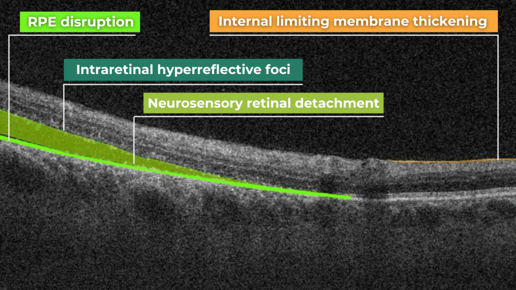
The software of the OCT machine automatically detects the border of the inner retina (internal limiting membrane) and the outer retina to calculate the retinal thickness map. The definition of outer retinal margin varies according to the imaging device (inner segment-outer segment junction for Stratus, inner part of RPE for Copernicus and Topcon 3D-OCT 1000, middle of RPE for Cirrus, outer part of RPE for Optovue RTVue 100, and Bruch’s membrane for Spectralis).
How OCT Works
OCT uses a low-coherence infrared light beam to scan the eye. The light that reflects back from different retinal layers is analyzed and converted into detailed images. This allows Dr. Rajeev Sudan and his team to assess the condition of the retina, optic nerve, and macula with incredible precision—often before symptoms are noticeable to the patient.
Key Components of the OCT System
Light Source: A broadband light source provides high resolution and deeper penetration.
Interferometer: Uses Michelson interferometry to detect delays between reflected beams.
Detector: Captures the interference pattern, which is then reconstructed into a clear digital image.
Applications of OCT
OCT is routinely used by Dr. Rajeev Sudan for diagnosing and managing a range of eye conditions, including:
Diabetic Retinopathy
Macular Degeneration
Glaucoma
Retinal Vein Occlusions
Macular Holes and Edema
The technology aids in detecting structural changes even in the earliest stages of disease, enabling timely treatment and better outcomes.
Why We Trust OCT at Dr. Rajeev Sudan’s Eye Clinic
High-Resolution Imaging: Micron-level detail allows for early diagnosis.
Non-Invasive & Painless: The scan takes just a few minutes and involves no discomfort.
Real-Time Results: Immediate visualization helps speed up diagnosis and treatment planning.
Our Commitment to Advanced Eye Care
By integrating Optical Coherence Tomography into routine clinical practice, Dr. Rajeev Sudan’s Eye Clinic Delhi, stands at the forefront of modern ophthalmic diagnostics. We are committed to delivering personalized, precision-driven eye care using the latest technology.
Whether you are managing a chronic condition like diabetes or simply undergoing a routine eye exam, OCT allows us to safeguard your vision with the utmost care and accuracy.

About Us
Dr. Rajeev Sudan, MD (AIIMS) is a cataract, glaucoma and refractive surgeon, with experience of more than 20 years. He is a post graduate and senior resident from Dr. RP centre, AIIMS and has received a honour of best resident award for an excellence in patient care at AIIMS. He has done more than 15000 surgeries including complex cataract , refractive and glaucoma surgeries.
Quick Links
Contact
A 6, Outer Ring Rd, near Savitri Flyover, Block A, Chittaranjan Park, New Delhi, Delhi 110019
rrsudan20@gmail.com
+91- 9958952302